
Computed Tomography (CT)
CT provides thin, cross-sectional images which appear as “slices.” These slices are then stacked together to create a three-dimensional image. By using x-rays to produce multiple images, the tool gives our team more detail than conventional radiographs. CT imaging is especially useful because it allows for views of all types of tissue, clearly showing bone, muscle and blood vessels.
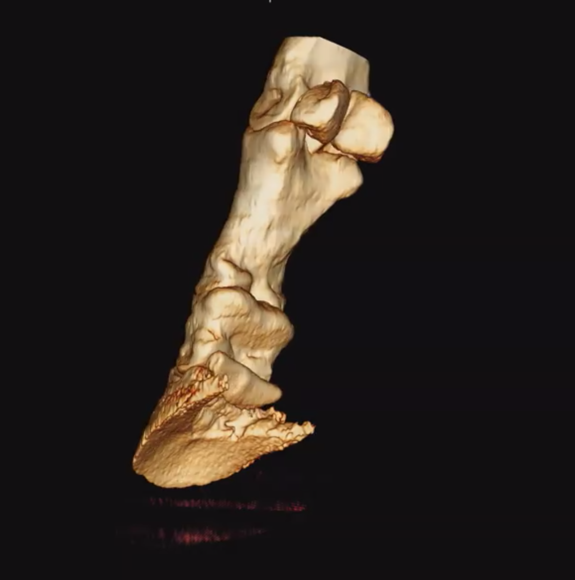
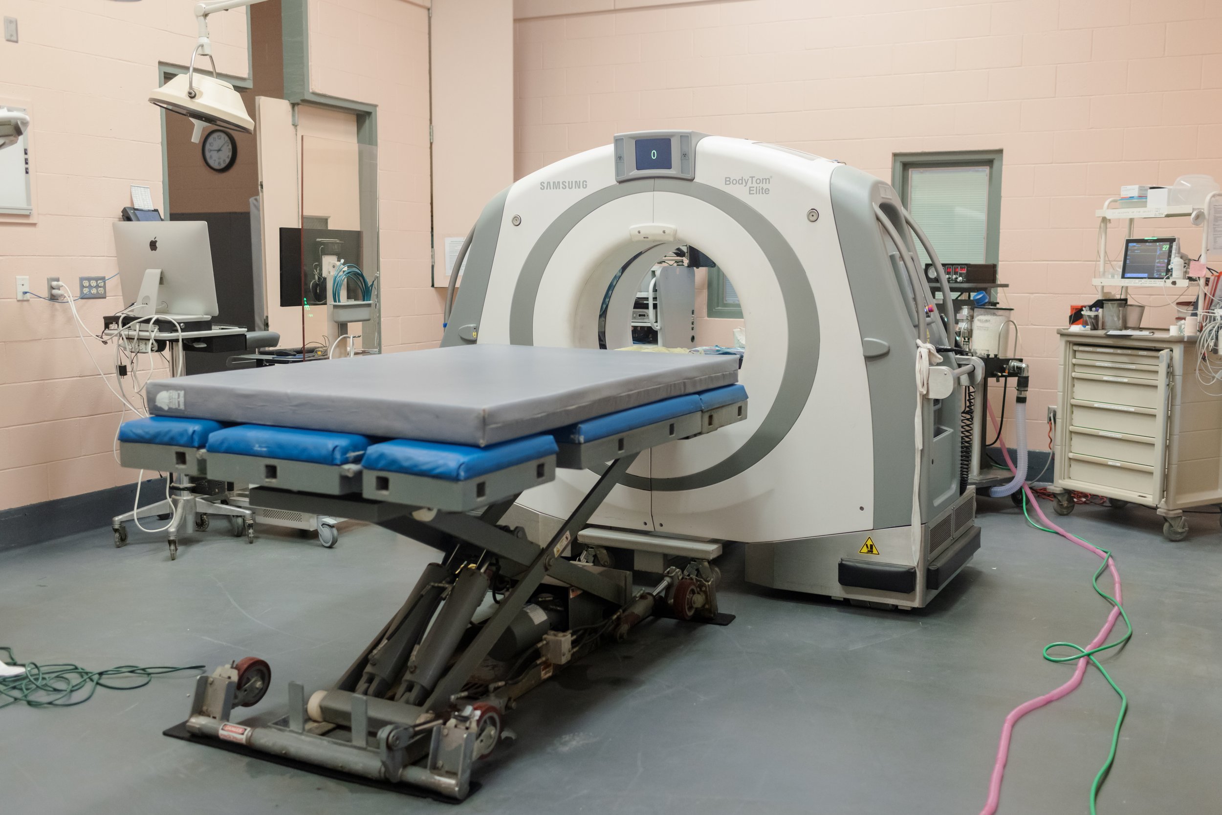
One of only three units of its kind in being used in equine veterinary medicine in the US, our team utilizes this advanced portable BodyTom® full-body, 64-slice CT to accurately explore issues within the equine head and neck and to examine complicated dental issues. This CT unit is also used to analyze fractures and other injuries to bones and joints of the distal limbs and to assist in the planning of complex surgeries.
“Having access to the most cutting edge technologies and tools available, means getting the most definitive diagnosis for the horse. ”